If you are working in a dental laboratory and have just bought a dental lab scanner, read on to find out how to ensure proper fitting for crowns! In this article, we will be sharing tips on scanning impressions, preparing the model, setting up the scan as well as which areas are the most important to scan correctly for that accurate fit.
With digital scanning, you can choose not to create a gypsum model from the impression you receive from a dental clinic and instead design the prosthetics based on the digital scan data itself. Whether you’d like to eliminate the model-making step or still incorporate it, here are tips for both cases.
Impression Scan
As we will be only using the scan data to make the prosthetics in this case, it is important to make sure that the impression has been done properly prior to scanning in order to design a properly fitted crown.
Firstly, check if there are issues with the impression due to saliva or blood on the abutment and contact point. If you scan an impression where the abutment is covered with saliva or blood, the model created using that impression will be inaccurate due to the difference in the abutment shape and the margin line.
Before scanning the impression, make sure that the impression itself has been made in the perfect condition for the prosthetics preparation. Once you’ve ascertained that the impression has no issues, you can begin the scanning process.
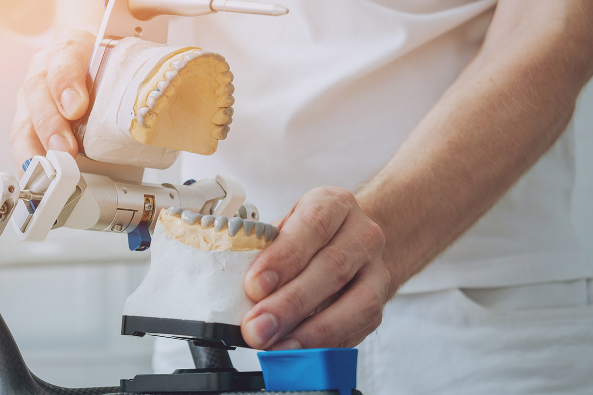
Model preparation
Before scanning the model, make sure that there is no undercut on the abutment teeth and that no defect had occurred during the impression pouring. In the case that there is an undercut in the abutment, block-out should be done using wax. This ensures a uniform thickness which is important in order to manufacture an accurate prosthetic.
If there is an undercut in the model itself, you will be unable to ensure uniform thickness which will cause issues with the manufactured prosthetics. The reason for ensuring uniform thickness is that the prosthetic manufactured needs to be made thick enough to withstand occlusal pressure during the manufacturing process. Hence, if there is an undercut, block-out operation should be done before scanning the model.
There is virtually no difference between scanning an impression directly or scanning a gypsum model created from an impression. Although it is possible to reduce errors while creating the gypsum model, it is important to know which parts are needed to create the prosthetics before scanning. This is because as long as these parts in the model are properly created, the measurements of the various elements can be adjusted accordingly in the CAD program itself.
Hence, the most important step in making accurate prosthetics would be firstly confirming the impression or model is accurate. This should be checked meticulously as you would check the scan data. Since both lab scanners and intraoral scanners are used to digitally fabricate prosthetics, it is imperative that the instruments being used are accurate and that the person operating these instruments have the required knowledge and skills to avoid errors.
We hope that you will be able to implement these tips to acquire more accurate scan data so that you can design and manufacture better-fitted crowns! Do subscribe to our blog for more of such tips on various applications for your lab scanners.