axsys, axis dental, axis dental milling, versamill, technology driven dental solutions, axis milling, top 5 milling machines in the dental industry, axsys technologies, axsysus, axsys inc, dental mill, axsys dental
The Future of Dental Technology: Trends to Watch
The dental industry is undergoing rapid transformation driven by technological advancements. As practices adopt digital solutions, staying informed about emerging trends is crucial for maintaining a competitive edge. Innovations such as artificial intelligence in diagnostics, tele-dentistry, and enhanced imaging technologies are reshaping patient care and operational efficiency.
For instance, AI-driven tools are enabling more accurate diagnostics and treatment planning, while tele-dentistry is improving access to care for patients in remote areas. Keeping abreast of these trends not only enhances service offerings but also positions dental practices as leaders in a fast-evolving market.
Maximizing ROI with Dental Equipment: A Strategic Approach
Investing in dental equipment requires careful consideration to maximize return on investment (ROI). Practices must evaluate not only the initial costs but also the long-term benefits of technology integration. A strategic approach involves assessing equipment performance, maintenance costs, and potential revenue generation from enhanced services.
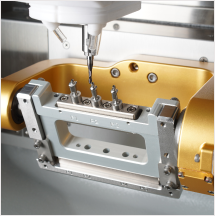
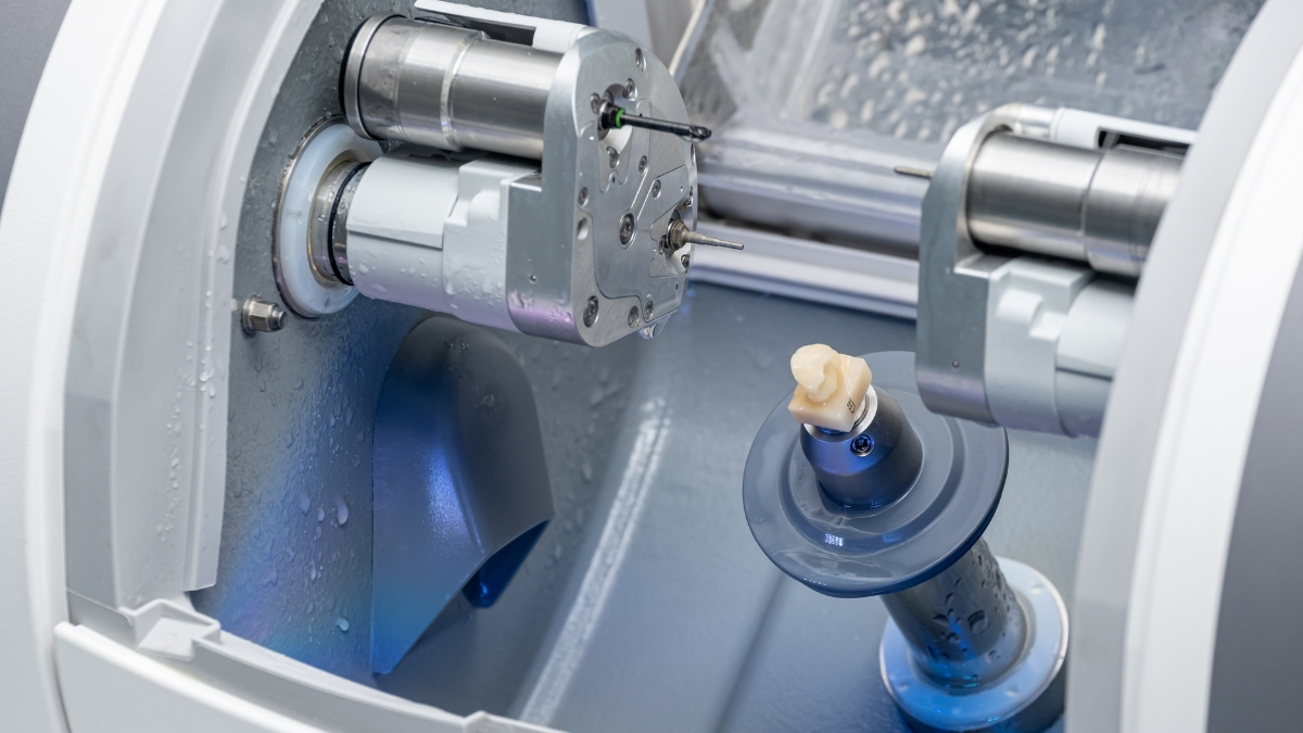
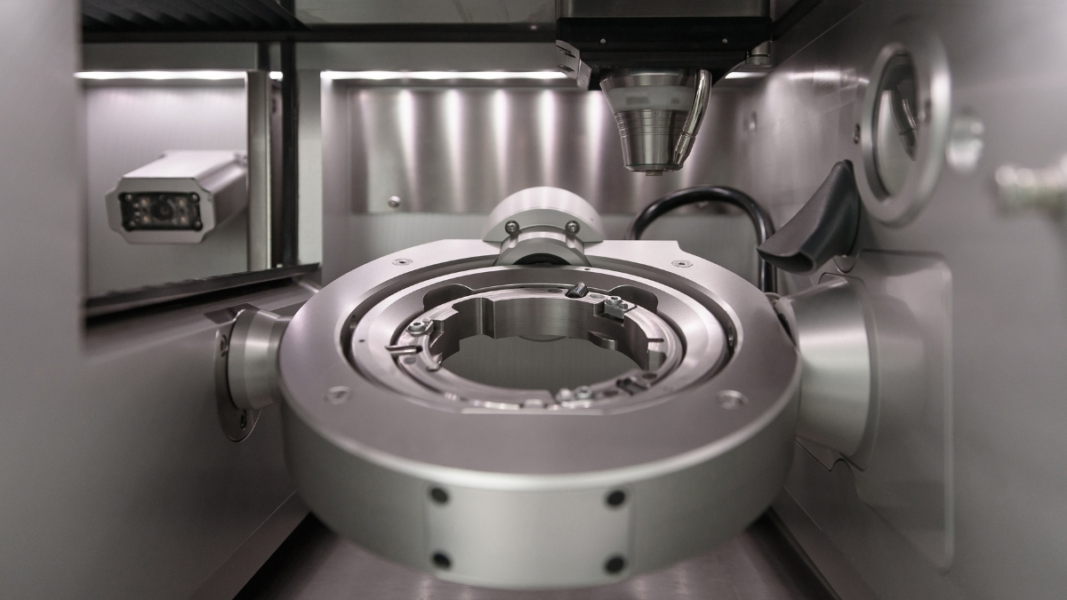
For example, upgrading to a high-precision milling machine can reduce production time and increase output, directly impacting profitability. Additionally, training staff to utilize new technologies effectively can lead to improved patient outcomes and satisfaction, further driving practice growth.
Building a Sustainable Dental Practice: Eco-Friendly Solutions
As the dental industry becomes more aware of its environmental impact, integrating eco-friendly solutions is gaining traction. Sustainable practices not only appeal to environmentally conscious patients but can also reduce operational costs. Implementing green initiatives such as digital workflows and energy-efficient equipment can significantly lower a practice's carbon footprint.
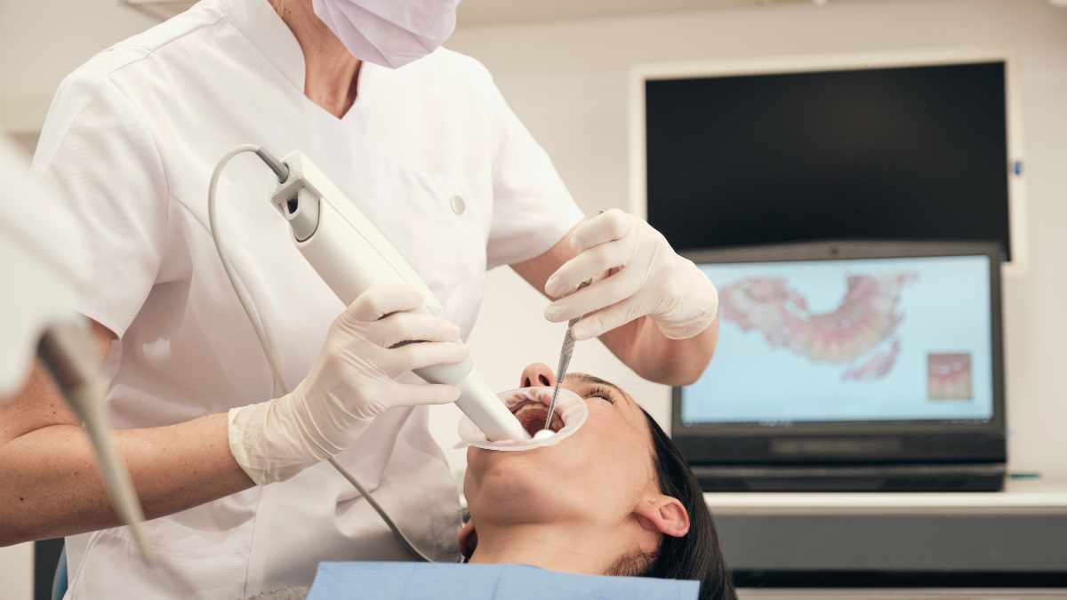
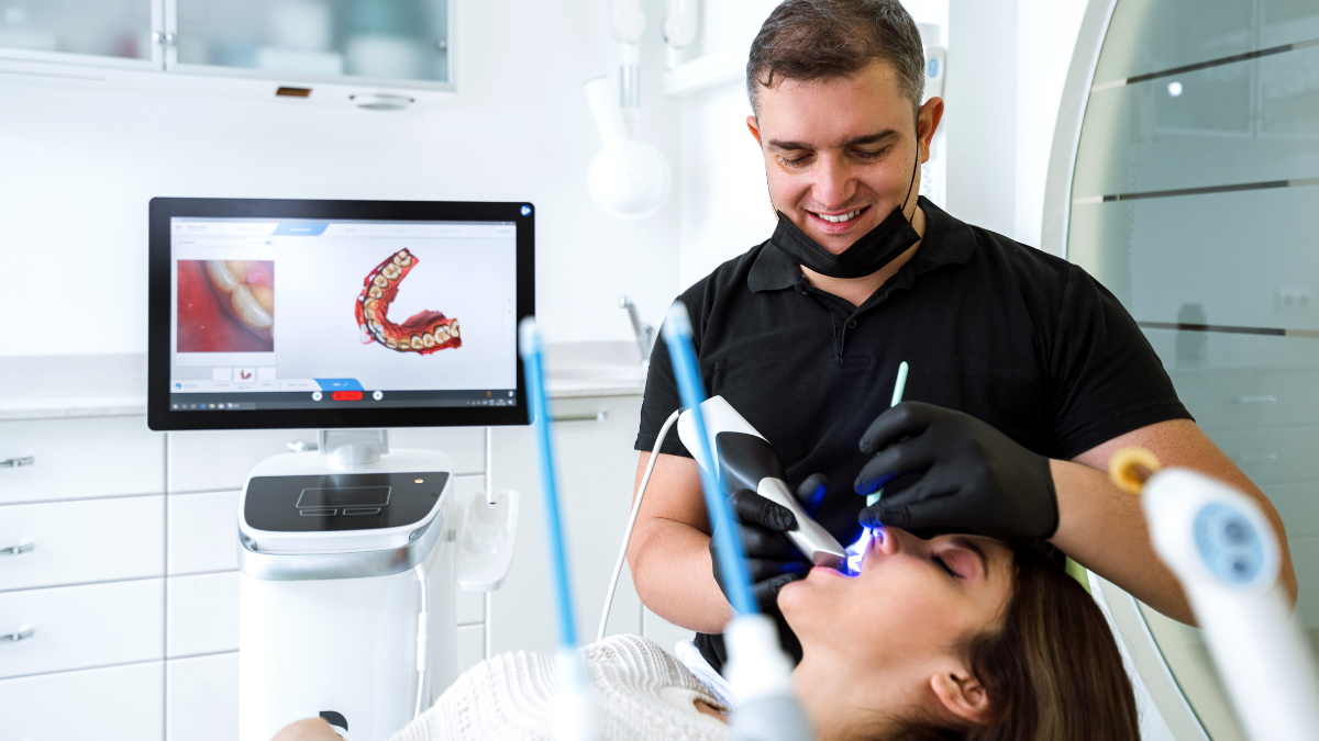
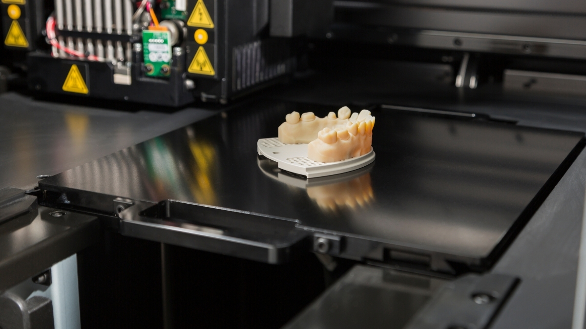
For instance, utilizing digital impressions reduces the need for physical materials, while energy-efficient 3D printers can minimize energy consumption. By adopting these practices, dental professionals can contribute to a healthier planet while enhancing their brand reputation and attracting a broader patient base.
Patient Education and Engagement: Enhancing the Dental Experience
Patient education is a vital component of modern dental care, fostering engagement and improving treatment outcomes. By providing comprehensive information about procedures and technologies, practices can empower patients to make informed decisions regarding their oral health. This engagement can lead to higher satisfaction rates and increased loyalty.
Utilizing various channels such as newsletters, social media, and in-office materials can enhance communication. For example, offering webinars on new technologies or treatment options can demystify dental procedures and create a more trusting relationship between patients and providers.


