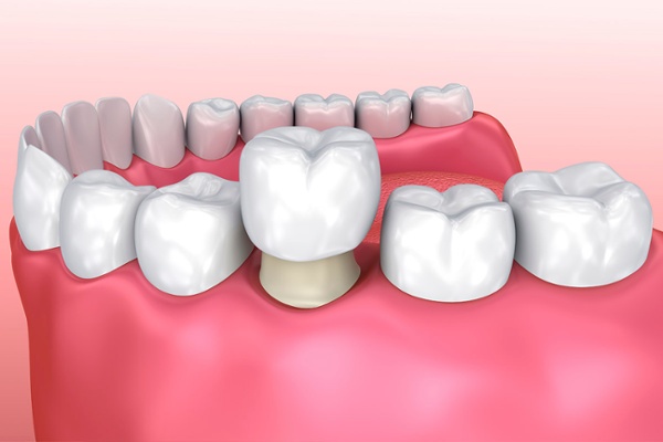
Now that you’ve bought an intraoral scanner, how do you use it to ensure the best results for your patients’ dental treatments? To help you get the best results with your intraoral scanner, we will be sharing tips on the proper usage of your scanner for various applications. For a start, here are some tips you can take note of during the scanning process to ensure that the resulting dental crown is properly fitted.
In order to design a properly fitted crown, the most important thing that you will require is accurate scan data. That is why you should always make sure that you are using the proper partial retraction method, and check that the scanner is capturing the impressions clearly, before starting the actual scanning. In addition, during a full arch scan, it is easy for errors to occur when scanning the region between the canines and premolar. Hence, you should spend more time scanning the regions at the corners of the mouth to capture more images to ensure more accurate data. The scan regions which need to be captured well in order to design a well-fitted crown are the working teeth, adjacent teeth, antagonistic teeth and bite.
Now, let’s take a look at specific tips to ensure quality scan data is captured during scanning.
Restoration Tooth (Teeth) Scan
Preparation Teeth: Before scanning, check whether there is an under cut and align the position according to the margin level.
If the patient has a supragingival margin, you only need to ensure proper fluid control (Saliva, Bleeding, Tissue Fluid) before scanning.
However, in the case of equigingival or subgingival margin, scanning without the use of cord packing would cause the margin and the gingiva to be out of alignment, resulting in an inability to acquire clear margin data. Hence, make sure to use cord packing when the margin is equigingival or subgingival, in order to acquire clear margin data.
Implant Case: You don’t need to take extra precautions when scanning the natural teeth. However, when scanning the area for the scan body, you should start scanning from the gingiva in order to avoid errors in the scan body data and ensure that its location is accurately captured.
Adjacent Teeth Scan
After reviewing the contact area of the restoration, you should make sure to properly scan the contact point areas as well as the areas which will impact the emergency profile.
Anterior Tooth: Aesthetics is important as it impacts many things. In order to get the exact tooth contour, you shouldn’t just scan the adjacent tooth but also the same name tooth along with the abutment tooth. In addition, to check occlusal relation, you should scan all the way to the premolar tooth.
Posterior Tooth: For posterior tooth, the mastication and occlusal relation are the most important factors, as opposed to aesthetics. Hence, in order to check the arch of the patient’s as well as his/her occlusal relation, you should scan the teeth at 1.5~2x the mesial/distal area of the abutment tooth.
Implant Case: Although the method of scanning is the same as when you scan natural teeth, when scanning implants, the emergency profile is more important than in abutment tooth cases. Hence, when scanning the gingiva, you should make sure to get a wide scan of the gingiva around the implant of the mesial/distal and buccal/lingual.
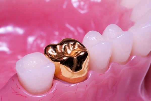
Gold Crown Case
The method of scanning is the same as when scanning normal adjacent teeth. However, you should consider using scan spray when scanning gold crowns as this makes scanning easier. When using scan spray, you will need to take into consideration the thickness of the scan spray powder as it could affect the accuracy of the scan. Adding too much powder may occur thickness between restoration and tooth and then affect the scan quality. Not to worry though! You can always use a suction to adjust the amount of the scan spray powder if you’ve applied too much.
We hope that these tips will enable you to acquire more accurate scan data in order to make better-fitted crowns! Do subscribe to our blog for more of such tips on various applications for your intraoral scanners.